Gene Delivery

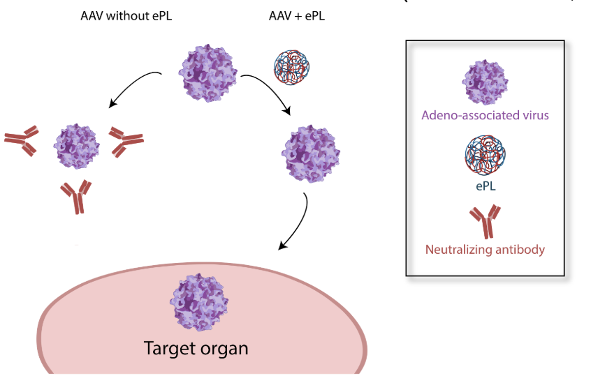
Development of gene therapy began about 50 years ago and has been used to cure numerous diseases, with some products reaching commercial use for a variety of diseases. The way gene therapy works is that a delivery vehicle carries a gene encoding for a healthy protein, and delivers that gene to cells in a patient with a mutated form of that gene. The patient’s mutated gene is what leads to their specific disease, either by failing to make a certain protein, or making that protein incorrectly. The healthy gene delivered by gene therapy can be read by the patient’s intracellular machinery and results in a functional protein, thus ameliorating patient symptoms. There are several delivery vehicles that are used in gene therapy, including viral vehicles, polymeric nanoparticles, and lipid nanoparticles. Our lab has interest in a range of these vehicles, but most of our work to date has been utilizing the viral delivery vehicle adeno-associated virus (AAV). We have engineered nanoparticles to be used in conjunction with AAV to increase its targeting in the body (thus enabling a lower dose) and to decrease its immunogenicity. (Switala et al 2024, J. Nanobiotechnology)
We Published These!
- Switala L, Di L, Gao H, Asase C, Klos M, Rengasamy P, Fedyukina D, Maiseyeu A. Engineered nanoparticles promote cardiac tropism of AAV vectors. J Nanobiotechnology. 2024 May 3;22(1):223.
- Mog B, Asase C, Chaplin A, Gao H, Rajagopalan S, Maiseyeu A. Nano-Antagonist Alleviates Inflammation and Allows for MRI of Atherosclerosis. Nanotheranostics. 2019 Nov 1;3(4):342-355.
